
Wydawcy
Katalog tematyczny
- Archeologia (109)
- Ekonomia, finanse, business (13704)
- Filozofia (1445)
- Historia (3040)
- Języki i językoznawstwo (2356)
- Lifestyle, hobby i rozrywka (475)
- Medycyna (9704)
- Nauki humanistyczne (4)
- Nauki o Ziemi i środowisku (2638)
- Nauki ścisłe (20412)
- Nauki społeczne (8486)
- Nauki techniczne (13450)
- Prawo (1696)
- Publikacje informacyjne i interdyscyplinarne (1288)
- Religia i wierzenia (917)
- Technika komputerowa (4067)
- Zdrowie, relacje międzyludzkie i rozwój osobisty (657)


Informacje szczegółowe o książce
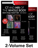
CT and MRI of the Whole Body, 2-Volume Set, 6th Edition
ISBN 9780323113281
Autor: John R. Haaga
Wydawca: Elsevier
Dostępność: 3-6 tygodni
Cena: 1 604,40 zł
Przed złożeniem zamówienia prosimy o kontakt mailowy celem potwierdzenia ceny.
ISBN13: |
9780323113281 |
ISBN10: |
0323113281 |
Autor: |
John R. Haaga |
Oprawa: |
Hardback |
Rok Wydania: |
2016-08-02 |
Numer Wydania: |
6 |
Ilość stron: |
2832 |
Now more streamlined and focused than ever before, the 6th edition of CT and MRI of the Whole Body is a definitive reference that provides you with an enhanced understanding of advances in CT and MR imaging, delivered by a new team of international associate editors. Perfect for radiologists who need a comprehensive reference while working on difficult cases, it presents a complete yet concise overview of imaging applications, findings, and interpretation in every anatomic area. The new edition of this classic reference - released in its 40th year in print - is a must-have resource, now brought fully up to date for today's radiology practice.
Review: <i>"Because it fits whole-body imaging into a succinct two-volume text, it will.make an excellent and often used addition to the library of general radiologists and radiologists in training.?</i> <b>Reviewed by</b> AJR, review of the previous edition <i>"CT and MR Imaging of the Whole Body maintains its status as a practical guide, combining cutting-edge research and theory into step-by-step descriptions of all CT and MR imaging applications.?</i> <b>Reviewed by</b> Radiology, review of the previous edition</p>
PART I PRINCIPLES OF COMPUTED TOMOGRAPHY AND MAGNETIC RESONANCE IMAGING Computed Tomography 1. Imaging Principles in Computed Tomography 2. Computed Tomography Imaging Operation Magnetic Resonance Imaging 3. Imaging Principles in Magnetic Resonance Imaging 4. Imaging Principles in Magnetic Resonance Angiography 5. Contrast-Enhanced Magnetic Resonance Imaging 6. Magnetic Resonance Imaging in the Pediatric Patient 7. Tissue Characterization in Liver Imaging Using Advanced MR Techniques PART II CT AND MR IMAGING OF THE WHOLE BODY Neuroradiological Imaging of the Brain and Meninges 8. Normal Anatomy 9. Intracranial Neoplasms 10. Cerebral Infections and Inflammation 11. Stroke 12. Cerebral Aneurysms and Cerebrovascular Malformations 13. Traumatic Brain Injury 14. Spinal Cord Injury 15. Neurodegenerative Disorders 16. Functional Magnetic Resonance Imaging 17. Brain Proton Magnetic Resonance Spectroscopy 18. Meningeal Processes 19. Demyelinating Disease and Leukoencephalopathies Neuroradiological Imaging of the Head and Neck 20. Orbit 21. Temporal Bone 22. Pharynx 23. Paranasal Sinuses 24. Cervical Adenopathy and Neck Masses 25. Larynx 26. Imaging of the Head and Neck in the Pediatric Patient Neuroradiological Imaging of the Spine 27. Non-Infectious Inflammatory Diseases Affecting the Spinal Cord 28. Spinal Trauma 29. Degenerative Disease 30. Spinal Tumors 31. Spinal Infections 32. Cystic Lesions 33. Spinal Vascular Diseases 34. Systemic Diseases Affecting the Spine 35. Congenital Abnormalities Imaging of the Chest 36. Non-neoplastic Parenchymal Lung Disease 37. Neoplastic Disease of the Lung 38. Mediastinal Disease 39. Disease of the Pleura, Chest Wall, and Diaphragm 40. Airway 41. Chest Imaging in the Pediatric Patient Imaging of the Abdominal and Pelvic Organs 42. Biliary Tract and Gallbladder 43. Liver: Normal Anatomy, Imaging Techniques, and Diffuse Diseases 44. Liver: Focal Hepatic Mass Lesions 45. Liver Transplantation 46. Pancreas 47. Mesentery 48. Spleen 49. Peritoneum 50. Gastrointestinal Tract 51. Rectum 52. Contrast Nephropathy and Its Management 53. Adrenal Glands 54. Kidney 55. Retroperitoneum 56. Male Pelvis 57. Female Pelvis Imaging of the Cardiovascular System 58. Coronary Arteries, Heart, and Pericardium 59. Advanced Cardiovascular CT Imaging Imaging of the Musculoskeletal System 60. Musculoskeletal Tumors 61. Shoulder 62. Hip and Pelvis 63. Knee 64. Foot and Ankle 65. High Resolution 3T Magnetic Resonance Neurography: Applications, Techniques, Pitfalls PART III IMAGE-GUIDED PROCEDURES (Frank K. Wacker, Part Editor) 66. MRI-Guided Interventions 67. Image-Guided Aspirations and Biopsies 68. Computed Tomography-Guided Drainage 69. Image-Guided Ablation of Parenchymal Organs PART IV LEADING EDGE IMAGING CONCEPTS 70. Vasculogenesis
Koszyk
Książek w koszyku: 0 szt.
Wartość zakupów: 0,00 zł
Informacje


Kontakt
Gambit
Centrum Oprogramowania
i Szkoleń Sp. z o.o.
Al. Pokoju 29b/22-24
31-564 Kraków
Siedziba Księgarni
ul. Kordylewskiego 1
31-542 Kraków
+48 12 410 5991
+48 12 410 5987
+48 12 410 5989
Subskrypcje
Administratorem danych osobowych jest firma Gambit COiS Sp. z o.o. Na podany adres będzie wysyłany wyłącznie biuletyn informacyjny.
Autoryzacja płatności
Informacje na temat autoryzacji płatności poprzez PayU.

© Copyright 2012: GAMBIT COiS Sp. z o.o. Wszelkie prawa zastrzeżone.
Projekt i wykonanie: Alchemia Studio Reklamy